
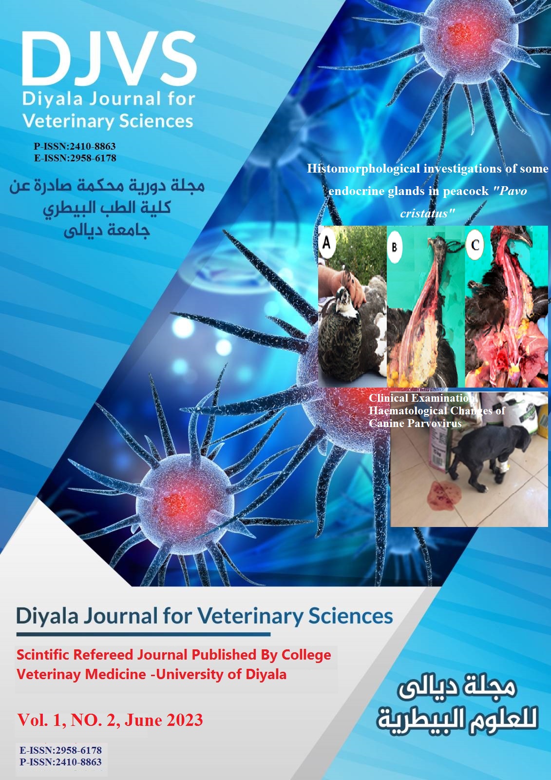
Clinical Examination, Haematological Changes of Canine Parvovirus with Laboratory Detection by Rapid Antigenic Test
DOI:
https://doi.org/10.71375/djvs.2023.01201Keywords:
Canine parvovirus, hematology, ImmunochromatographyAbstract
Background: Cases of canine parvovirus have increased in the last few years in Iraq.
Aims: to identify clinical, haematological changes in dogs infected with canine parvovirus
Material and Methods: A total of 50 dogs of various ages, sexes, and breeds were clinically investigated from December- February2021. Sterile swabs were used for collection of fecal samples. CPV using a commercially available quick CPV antigen detection test kit.
Results: Only 40 dogs were infected with CPV, which is clinically exhibited by enteric form lethargy, weight loss, lack of appetite, diarrhea, and develops to cause blood-tinged or bloody diarrhea, foul-smelling vomiting, and intractable fluidy diarrhea. Hematological analysis of the samples with neutropenia, lymphopenia, and monocytopenia revealed statistically significant declines (P 0.05) in the RBCs, Hb, PCV, MCV, MCH, MCHC, and WBCC. Anomalies in erythrocyte morphology included leptocytes, echinocytes, schistocytes, hypochromia, anisocytosis, and poikilocytosis. Infection with CPV more commonly among males (81.5%) than female (78.3%). CPV infection more commonly in younger ages a dog. There is a correlation between infection rates and breeds of dogs, with German shepherds and Terriers having higher infection rates (88.2% and 85.0%, respectively) than other breeds (57.1%).
Conclusions: CPV infect younger age, male dogs with significant hematological changes
Downloads
Downloads
Published
How to Cite
Issue
Section
License
Copyright (c) 2023 Tareq Rifaaht Minnat, Zahra Jafaher Sadiq

This work is licensed under a Creative Commons Attribution-NonCommercial 4.0 International License.

